Skinic
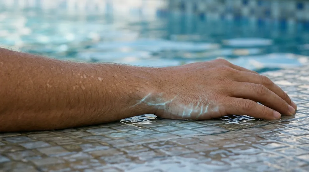
White spots on skin come from five common causes: a yeast that returns each summer, ageing and sun, a rash or procedure that has healed, dry patches in children, and vitiligo. They are told apart less by colour than by where the spot sits, whether it has scale and when it appears. Only the last of the five is a condition to have looked at rather than watched.
The claim this article tests is that white spots mean a calcium deficiency. It covers each cause in turn, why the colour can outlast the cause, and which patches deserve a clinician. It describes and evaluates. It does not diagnose a patch or direct treatment, and a patch you are unsure about is a question for your own clinician. Raised white bumps and white spots on the lips are different problems, covered in our articles on milia and white spots on lips.
Five Causes, Three Questions
Pale patches look alike from a distance and differ up close. The table sets out what the sources describe for each cause.
| Cause | Look | Usual site | Scale | Who, and when |
|---|---|---|---|---|
| Pityriasis versicolor | Oval patches, pale, pink or brown, that can merge | Trunk, neck, upper arms | Fine scale | Teens and young adults; warm, humid months |
| Idiopathic guttate hypomelanosis | Flat dots, 2 to 5 mm | Forearms, shins, V of the chest | Smooth in the usual form | Becomes more common with age |
| After inflammation | Pale area where skin was inflamed, burned or treated | Wherever the cause was | Depends on the cause | After the cause has passed |
| Pityriasis alba | 1 to 20 poorly defined patches, 0.5 to 5 cm | Cheeks and chin | Fine scale at one stage | Children aged 3 to 16 |
| Vitiligo | Milky-white, well-defined patches | Often symmetrical: hands, around eyes and mouth, skin creases | Not described as scaly | Any age; average onset 20 to 24 |
Where Is It?
Location narrows the field and does not settle it. DermNet places pityriasis versicolor on the trunk, neck and arms, and calls it uncommon elsewhere. Its page on idiopathic guttate hypomelanosis names the sun-exposed forearms, shins and V of the chest. Pityriasis alba favours the cheeks and chin. Vitiligo can appear anywhere but is more common around the eyes and lips, on fingertips and in body folds, and at sites of injury.
Is There Scale?
Fine scale points to pityriasis versicolor or pityriasis alba, and a smooth surface to the other three. Scale can be hard to see. A 2022 review notes that the scale of pityriasis versicolor is often difficult to appreciate until the skin is stretched or scraped, and that treated or burned-out patches usually lack it. DermNet adds that a patch can begin scaly and brown, then pass through a non-scaly white stage. A smooth patch therefore does not rule out the yeast.
What Time of Year, and What Age?
DermNet says pityriasis versicolor most often affects young adults and may clear in winter and recur each summer. Pityriasis alba is a condition of children and adolescents, and its patches look paler in summer because tanning of the surrounding skin makes them more prominent. Idiopathic guttate hypomelanosis becomes more common with age, affecting 90% of people aged 81 to 90. For vitiligo, the average age of onset is 20 to 24 and 80% of cases appear before 30, and light-skinned people tend to notice it in summer when the contrast with tanned skin sharpens.
Fungal Patches That Come Back Each Summer
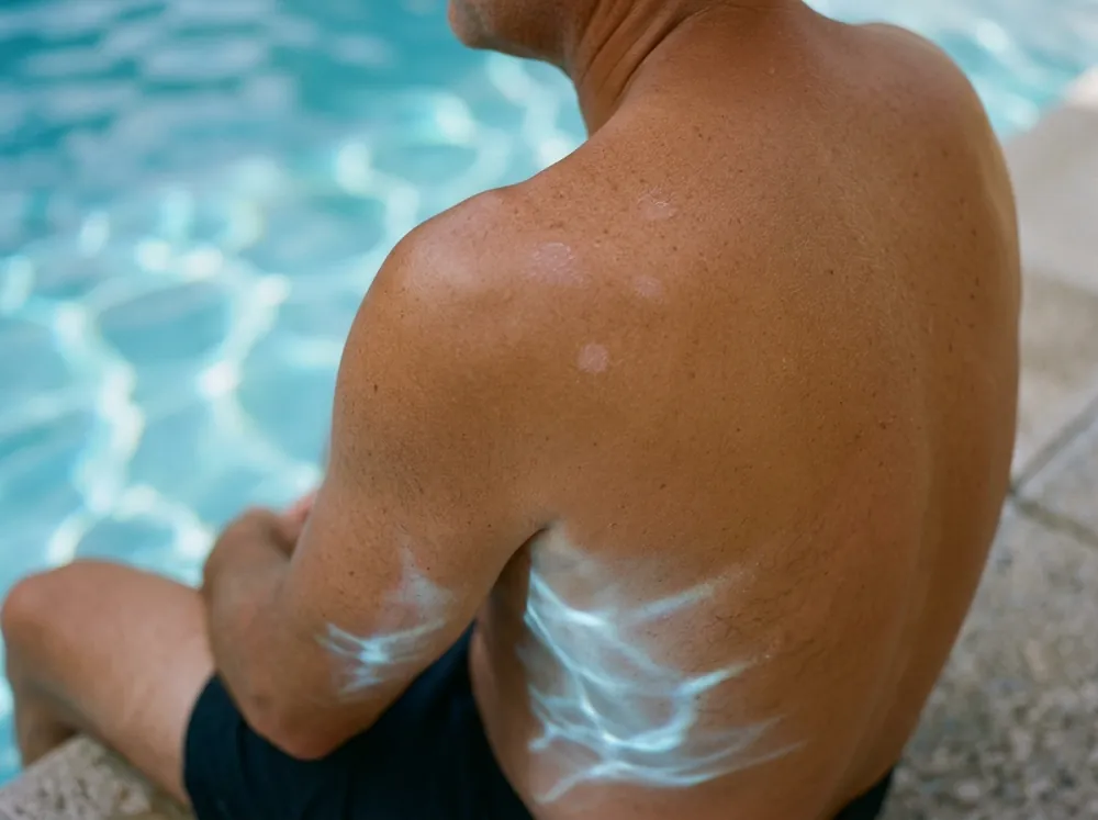
Pityriasis versicolor is a common yeast infection of the skin, caused by Malassezia. These yeasts are part of normal skin and depend on oil to live, and DermNet says it is not known why they sometimes grow actively enough to form patches. The review lists hot, humid weather, heavy sweating, oily lotions and creams and mask wearing among the predisposing factors. This is the "fungus" in fungus white patches on skin. DermNet notes that the older name tinea versicolor is used loosely, because tinea strictly means a dermatophyte infection, which this is not.
The pale type is thought to follow a chemical made by the yeast that impairs the pigment cells, and the review points to azelaic acid. Patches may be coppery brown, paler than the surrounding skin or pink, and pale patches are more common in darker skin. DermNet says the condition is not infectious in the conventional sense, although more than one family member is sometimes affected.
The colour can outlast the yeast. DermNet states that the fine surface scale is the sign of active infection and that once it has gone the infection is no longer ongoing. The colour may persist for weeks or months, particularly the pale type, and does not indicate treatment failure. DermNet lists topical antifungals for mild disease and oral antifungals for extensive disease or when topical treatment has failed, and both sources say relapse is common because the yeast is a normal resident of skin. A clinician confirms the diagnosis, sometimes with a scraping or a lamp examination.
Small White Dots on the Shins and Forearms
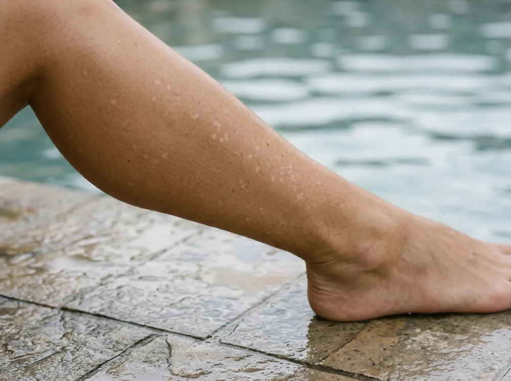
DermNet describes idiopathic guttate hypomelanosis as a benign, common, acquired condition with small flat pale or white spots on sun-exposed limbs. The spots are usually 2 to 5 mm across, occasionally up to 1.5 cm, on the forearms, shins and V of the chest. They are multiple and do not itch. That description fits many searches for tiny white spots on skin and for white spots on legs, though it is a description and not a diagnosis. The dark marks that appear on the same sun-exposed sites are covered in our article on sun spots, age spots and freckles.
The cause is not fully understood. DermNet calls it probably multifactorial, with skin ageing, chronic sun exposure, genetic factors and, less often, trauma or autoimmune factors, and reduced transfer of melanin from pigment cells to the skin cells around them. It affects all skin types and is more obvious in skin of colour.
The course is slow. DermNet states that it does not spontaneously repigment and that lesions may slowly enlarge and multiply with age. It usually needs no treatment beyond reassurance, with sun protection recommended. The page lists several procedural and topical options and gives outcome detail for only one, a single 5-second cryotherapy session with repigmentation visible by four months.
Pale Patches After Inflammation
Two different things sit here: skin that has healed after being inflamed or injured, and a childhood condition classed as a mild form of eczema.
After a Rash, Burn or Procedure
DermNet's leukoderma page separates leukoderma, a white patch from total loss of epidermal melanin, from hypopigmentation, which is paleness from reduced pigment. Its list of causes includes severe skin disease such as atopic dermatitis and allergic contact dermatitis, shingles, burns and other injuries, scarring, and procedures such as cryotherapy, chemical peels and laser. It is more obvious in tanned skin and skin of colour.
Recovery depends on the cause. DermNet says leukoderma may recover slowly if the cause can be avoided, such as stopping a drug or chemical exposure, or treated, such as an inflammatory skin condition. Leukoderma from scarring is likely to be stable and persistent. Our article on eczema on the face covers the rashes that can come first, and the opposite change, dark marks after inflammation, is covered in hyperpigmentation in brown and deep skin tones.
Dry Patches on a Child's Cheeks
DermNet describes pityriasis alba as a low-grade eczema seen mainly in children aged 3 to 16, with a prevalence around 5%. Typically there are 1 to 20 round, oval or irregular patches of 0.5 to 5 cm, usually on the cheeks and chin, with poorly defined edges and little or no itch. A patch passes through stages: a slightly scaly pink patch, a pale patch with fine scale, then a pale patch without scale, then resolution.
The cause is unknown. It often coexists with dry skin and atopic dermatitis, and DermNet says researchers have reached no conclusions about ultraviolet radiation, bathing habits, low copper and other trace elements, or Malassezia. No treatment is needed when it causes no symptoms, a moisturiser may improve the dry look, and the colour returns completely, after an average of one year with a range of a few months to two or three years.
When It Is Vitiligo
DermNet defines vitiligo as an acquired, chronic, depigmenting disorder in which pigment cells are progressively lost, appearing as milky-white patches. It states that autoimmune destruction of those cells is the widely accepted cause. It affects 0.5 to 2% of people. The NHS says vitiligo is not caused by infection and cannot be caught from someone else.
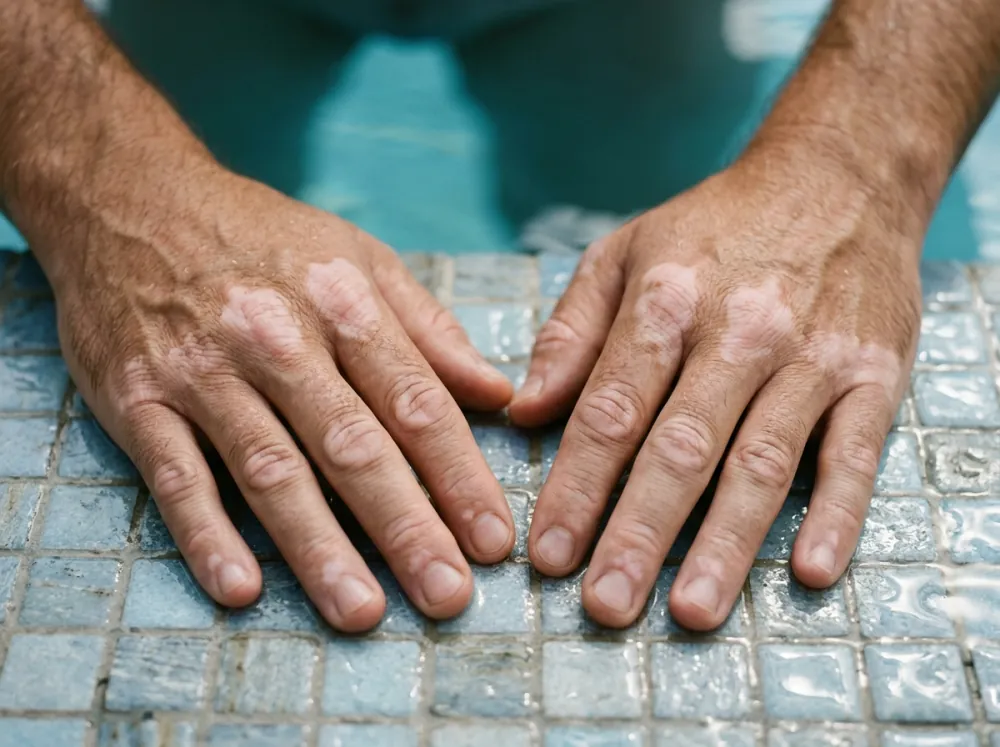
Several features separate it from the other four. The pigment loss is complete, so the patch is chalk- or milky-white and the surface is normal. It tends to be symmetrical and to favour sites that are rubbed or injured, a pattern DermNet calls the Koebner phenomenon. Hair in a patch can turn white. DermNet reports an association with thyroid disease, affecting up to 15% of adults with vitiligo, and the NHS says a blood test of thyroid function may be arranged.
Depigmented skin has no protection of its own. DermNet states that it can burn but cannot tan, and that sunburn may make vitiligo spread. The two sources differ on treatment. The NHS says colour restored by treatment does not usually last and that treatment cannot stop vitiligo spreading. DermNet says the aim is to stop progression and gain repigmentation, and describes a ruxolitinib cream approved by the FDA for non-segmental vitiligo from age 12 on the strength of two phase 3 trials, with body vitiligo responding less well than facial. It also says no measures are proven to prevent vitiligo, and that the vitamin supplements and alternative remedies endorsed by support groups are not based on scientific evidence.
The Figure Audited: "White Spots Mean a Calcium Deficiency"
Verdict: unsupported by any source opened. None names calcium as a cause of any of the five. Findings:
- The causes sections do not mention it. They name a yeast (pityriasis versicolor), ageing, chronic sun exposure and genetics (idiopathic guttate hypomelanosis), inflammation and injury (post-inflammatory leukoderma), an unknown cause (pityriasis alba), and autoimmune loss of pigment cells (vitiligo).
- Nutrition is addressed only to say it is unproven. DermNet lists vitamin deficiencies among factors described as precipitating vitiligo, and states that correlation is still not proven. For pityriasis alba it says no conclusions have been reached about low copper and other trace elements.
- Supplement remedies are not supported either. DermNet says those endorsed for vitiligo are not based on scientific evidence.
- The limit of this audit. Absence from five reference pages is not proof that no link exists, and white marks on the nails are a different question that these sources do not cover.
When a White Patch Needs a Clinician
None of the sources frames a self-test, and a patch cannot be sorted into a cause from a photograph. DermNet and the NHS both describe diagnosis as a clinical examination, sometimes with a Wood's lamp. These features fit the vitiligo pattern or another cause that a clinician should see:
- a milky-white, well-defined patch with a normal surface, especially if symmetrical or near the eyes, lips, hands or skin creases
- new patches, or existing ones that are spreading
- hair inside a patch turning white
- a white area inside or beside a mole. DermNet lists white regression areas within a melanoma, and white patches elsewhere in someone with melanoma, as melanoma-associated leukoderma. As reasoning, any white change near a mole belongs in front of a clinician
- a patch in a child that does not match pityriasis alba's pattern of cheeks, fine scale and little itch
A scaly patch that returns every summer is more likely the yeast, and a clinician can confirm that quickly. Because depigmented skin burns easily, sun protection matters while the patch is being assessed.
Conclusion
White spots on skin have five common causes, and the three questions of where, whether scaly and when sort them better than colour does. The yeast returns each summer and leaves colour behind for weeks. Small flat dots on the shins are usually idiopathic guttate hypomelanosis and arrive with age. Healed rashes and a child's dry cheeks leave pale skin that fades. Vitiligo is the one with a pattern worth showing to a clinician. No source opened links any of them to calcium.
If a patch is changing, photographing it in the same light each month, in an album or in an app like Skinic, gives a clinician a record of whether it has spread. For a patch you want identified, ask your own clinician.
Frequently asked questions
5 questions · tap one to open the answer
What causes white spots on the skin?
Are white patches on the skin a fungus?
Do white spots on the skin mean a calcium deficiency?
What are tiny white spots on the legs?
How is vitiligo different from other white patches?
Medically reviewed by
Dr. M.M Hanaei
Aesthetic Physician, Dermatology Fellowship
- MD — Shahid Beheshti University of Medical Sciences (SBMU)
- MCC Licensure (Medical Council of Canada)
- Dermatology Fellowship, Switzerland
- Certified in advanced aesthetic procedures
Last reviewed 16 October 2026