Skinic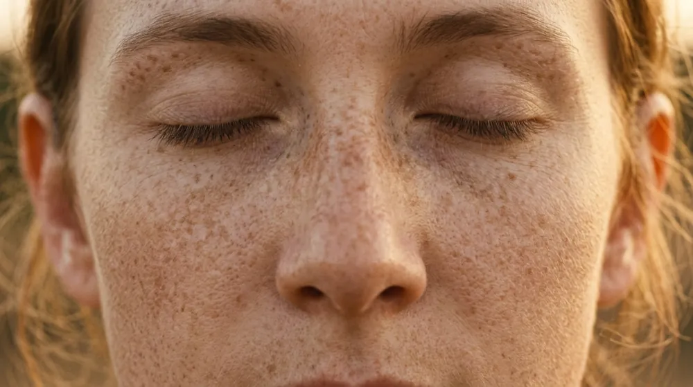
Sun spots, age spots and liver spots are three names for one thing, and freckles are a different one. DermNet lists sun spots, age spots, liver spots and "senile freckles" as common names for the solar lentigo, a benign flat pigmented mark caused by chronic ultraviolet exposure. A freckle is an ephelis, an inherited trait that darkens in summer and fades in winter.
That is the easy part. The harder part is that DermNet also lists a missed diagnosis of skin cancer, such as melanoma in situ, as a complication of solar lentigo, because the two can look alike. This article covers what separates freckles from lentigines, why age is not the cause, where each appears, the features that mean a spot is not a sun spot, and where to read about fading. It describes and sorts. It cannot tell you that a particular spot is harmless, and a spot you are unsure about belongs with a clinician.
Three Names, Two Things
The terms map like this.
| Common name | Clinical name | What it is |
|---|---|---|
| Sun spot, age spot, liver spot, senile freckle | Solar lentigo (plural lentigines) | A flat, benign pigmented mark from chronic UV exposure |
| Freckle | Ephelis (plural ephelides) | A small light brown or tan mark, inherited, from a variant MC1R gene |
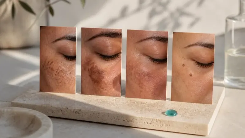
"Liver spot" has nothing to do with the liver. Our article on the types of dark spots on the face places both beside melasma and post-inflammatory marks.
Freckles Fade, Lentigines Stay
The seasonal test is the cleanest practical difference between sun spots and freckles, and DermNet states it on both pages.
| Freckle (ephelis) | Sun spot (solar lentigo) | |
|---|---|---|
| When it appears | Childhood; not present at birth | Mostly middle-aged and older adults |
| Edge | Poorly defined, may merge into patches | Well demarcated |
| Size | 3 to 10 mm | A few millimetres to centimetres |
| Colour | Light to dark brown | Yellow, tan, dark brown or black, typically uniform within each spot |
| Season | Prominent in summer, fade in winter | Persist year-round, may darken slightly in summer |
| Cause | Inherited MC1R variant; darkens with UV | Chronic UV exposure |
DermNet's freckle page adds that ephelides are common in fair-skinned Celtic children, that people with many have a variant of the MC1R gene, and that the number increases after UV exposure. A lentigo can feel rough or scaly to the touch because of thickened epidermis. Our article on sunscreen for hyperpigmentation covers what sunscreen does for each.
Why They Cluster Where They Do
Where Each Appears
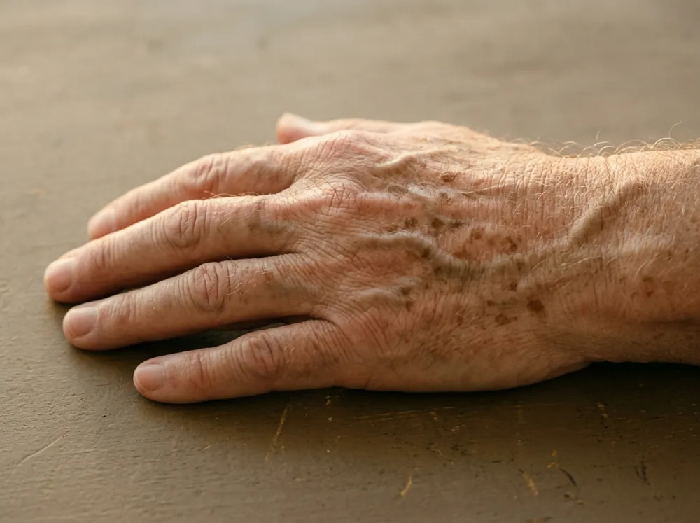
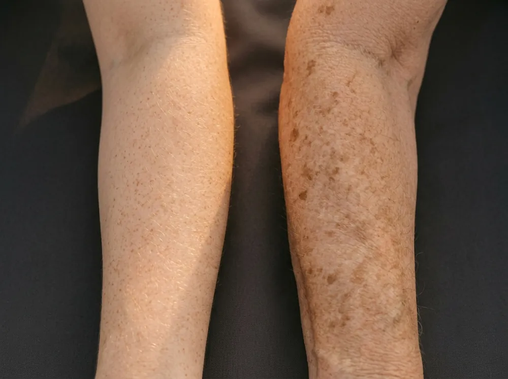
Both marks follow the sun. DermNet places freckles on sun-exposed sites, particularly the nose and cheeks, and solar lentigines on the backs of the hands, forearms, upper body and shins, and on the face in the Asian population. They are associated with lighter skin (Fitzpatrick phototypes I and II) and found in up to 90% of Caucasians over 60, but DermNet notes solar lentigo "may be more difficult to appreciate in darker skin types". On the face, sun spots and age spots on the face are the same solar lentigines, while freckles favour the nose and cheeks. Our article on Fitzpatrick skin types explains the scale, and the one on hyperpigmentation in deep skin tones covers how marks present there.
Why Clustering Matters
Clustering has a consequence beyond appearance. DermNet calls solar lentigines an independent risk factor for melanoma, and a 2020 melanoma risk model (abstract) that included clinically assessed naevi and upper-back solar lentigines found naevi the strongest predictor, with solar lentigines on the upper back also in the final model. The marks themselves are benign, and they record the UV exposure behind that risk.
The Figure Audited: "Age Spots Are Caused by Age"
Verdict: age is associated with age spots, and UV is the cause. Findings:
- DermNet states the cause is ultraviolet radiation, from the sun or artificial sources such as sunbeds and phototherapy, and says a lentigo can appear on sun-damaged skin after an acute sunburn.
- They are uncommon in young children unless sun exposure was significant, or the child has xeroderma pigmentosum, which fits exposure over time and not age as such.
- Other factors predict them. In a study of 523 French women (abstract), the severity of solar lentigines was associated with age and with five other factors: dark skin colour and tanning capacity, a history of freckles, sun-exposure behaviour, and use of oral contraceptive or progestogen treatments. The authors conclude lentigines are markers of photoaging, whereas freckles are mainly determined by genetics.
- The mechanism is not fully settled. DermNet says the exact pathogenesis is not fully understood, with UV thought to cause mutations that enhance melanin production and retention.
Two limits apply. The French study is cross-sectional and reports associations, and the hormonal finding, which the authors say merits further investigation, is not a settled cause. The name "age spot" is shorthand for cumulative UV exposure, which accumulates with age.
The two DermNet pages also differ slightly on melanocyte numbers: the solar lentigo page says they are normal or only slightly increased, while the freckle page says there is no increase in melanocytes, in contrast with lentigines. The difference is small, and both agree the marks involve more pigment, not a tumour.
The Spot That Is Not a Sun Spot
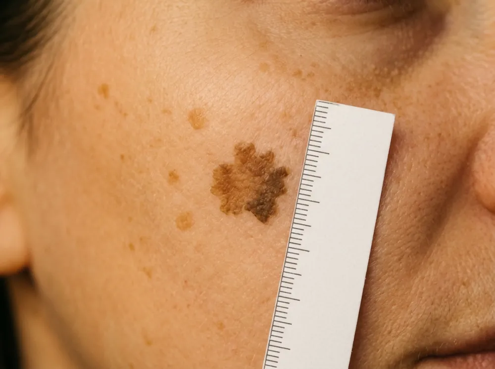
A solar lentigo is diagnosed clinically, helped by dermoscopy. DermNet's differential diagnosis for it includes atypical solar lentigo, macular seborrhoeic keratosis, pigmented actinic keratosis, pigmented Bowen disease, melanoma in situ, freckles and a junctional naevus. It says solar lentigines with unusual features that make them hard to tell from melanoma in situ are called atypical, that a biopsy is performed where there is diagnostic uncertainty, and that if a lesion is variegated in colour, biopsies come from each distinct area, since different conditions can sit within one spot.
The ABCDE Features
The American Academy of Dermatology's ABCDEs of melanoma are:
- A, asymmetry: one half of the spot is unlike the other.
- B, border: irregular, scalloped or poorly defined.
- C, colour: varying shades of tan, brown or black, or areas of white, red or blue.
- D, diameter: melanomas are usually larger than 6 millimetres, about a pencil eraser, when diagnosed, but can be smaller.
- E, evolving: the spot looks different from the rest or is changing in size, shape or colour.
The Academy says a new spot, a spot that is different from others, or one that is changing, itching or bleeding is a reason to see a board-certified dermatologist. DermNet's melanoma page restates the letters as A, B, C, "Different" (formerly diameter) and E, adds E, F, G (elevated, firm, growing) for nodular melanomas, and says any lesion that changes in size, shape, colour or elevation for more than one month should be reviewed by a dermatologist or biopsied. It cites a diameter above 7 mm in the Glasgow checklist, so thresholds vary between 6 and 7 mm. As reasoning, that variation is one reason size alone is a poor test.
Why Early Looks Matter
Lentigo maligna is the most common clinical subtype of late-onset melanoma, often on the head and neck, in accumulated sun exposure. DermNet's page on melanoma in situ says the cells are confined to the epidermis, that it has no metastatic potential in that stage, and that less than 5% of lentigo maligna is thought to become invasive, but that some in-situ melanomas develop foci of invasive melanoma and that detection at the in-situ stage improves survival.
In skin of colour DermNet notes melanomas can be harder to identify, tend to be thicker at diagnosis, and more often occur on the palms, soles and nails. A mark that is not on a sun-exposed site is not automatically a sun spot.
What Fades Them
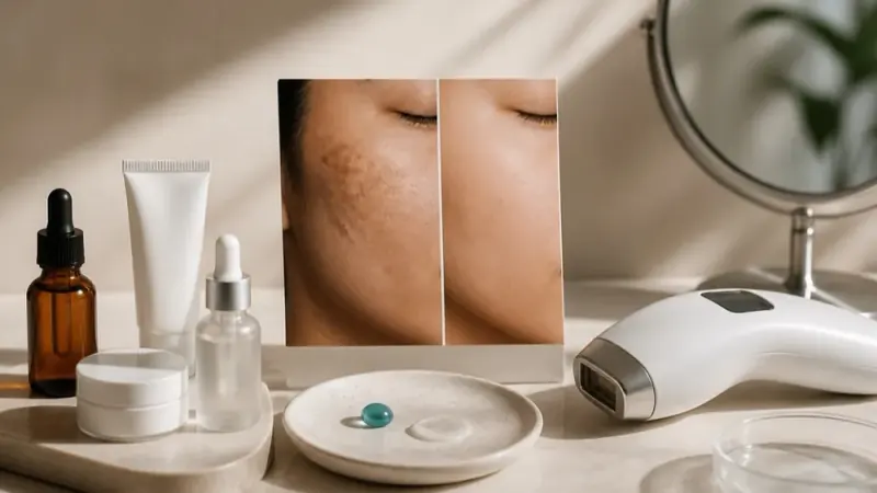
Fading is covered elsewhere, and the sources agree it is optional. DermNet says solar lentigines do not require treatment unless for cosmetic reasons, that sun protection reduces new ones and may modestly lighten existing ones, and that combination therapy has the highest resolution rates, with destructive methods such as cryotherapy and lasers carrying a risk of pale or dark spots. Our article on removing dark spots on the face covers the options.
As reasoning, not a finding, lightening or destroying a spot that has not been looked at removes the sample a clinician would examine, which is a reason to have an uncertain spot assessed first.
Conclusion
Sun spots, age spots and liver spots are solar lentigines, caused by cumulative UV exposure, and freckles are inherited marks that fade in winter. The seasonal test sorts them, and age is a correlate, not the cause. What a fingertip or a mirror cannot do is separate a lentigo from a lookalike, and DermNet lists missed melanoma in situ as a complication of exactly that confusion.
If you want a record of whether a spot is changing, photographing it with a ruler in the same place and light each month, in an album or in an app like Skinic, gives a clinician something to compare. For any spot that is new, different, changing, itchy or bleeding, ask your own clinician.
Frequently asked questions
5 questions · tap one to open the answer
What is the difference between sun spots, age spots and freckles?
Are age spots caused by age?
Do freckles and sun spots go away?
How can you tell if a brown spot might be melanoma?
Are sun spots a sign of skin cancer?
Medically reviewed by
Dr. M.M Hanaei
Aesthetic Physician, Dermatology Fellowship
- MD — Shahid Beheshti University of Medical Sciences (SBMU)
- MCC Licensure (Medical Council of Canada)
- Dermatology Fellowship, Switzerland
- Certified in advanced aesthetic procedures
Last reviewed 15 October 2026