Skinic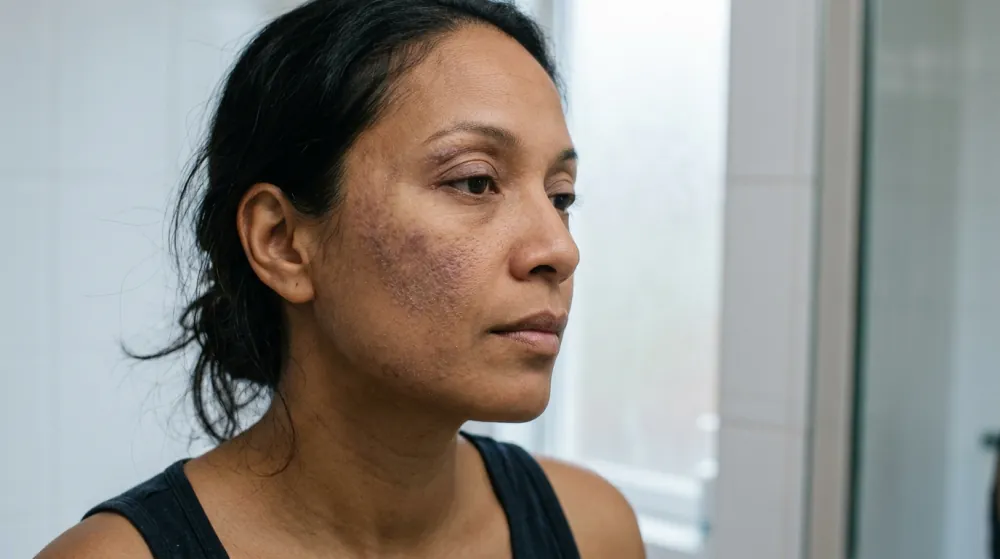
Eczema on the face is not one condition, and the commonest eyelid diagnosis in patch-test clinics is not the atopic kind. In a North American analysis of more than 50,000 patients referred for patch testing, allergic contact dermatitis was the final diagnosis in 43% of those with eyelid dermatitis only, against 13% for atopic dermatitis. Those patients were referred for patch testing, so the numbers describe a selected group.
On darker skin, the redness that eczema pictures show on pale skin can look violet, brown or grey. DermNet notes that this means scoring systems may underestimate severity, and that post-inflammatory colour change is more noticeable.
This article on eczema on the face covers where it shows, the atopic, contact and seborrheic types, why eyelids differ, the claim that facial eczema is always atopic, the triggers worth testing, why facial skin needs gentler handling and when to be seen. It describes and does not diagnose. A rash that is new, spreading, painful, blistering or not settling is a question for your own clinician.
Where It Shows on the Face
Eczema on face skin has patterns, and the sources describe them by type.
- Cheeks and around the mouth in infants. DermNet describes atopic dermatitis in babies as often beginning on the scalp and skin creases, and then involving the face, especially the cheeks.
- Eyelids and under the eyes. Adult atopic dermatitis can involve the eyelids, and DermNet lists a fold of skin under the lower eyelids (a Dennie-Morgan fold) and loss of eyebrow hair from rubbing as clues to atopy.
- Eyebrows, nasolabial folds, glabella, hairline and ears. DermNet lists these oil-rich sites for seborrheic dermatitis, with petal-shaped flaky patches at the hairline.
- The eyelids alone. Eyelid dermatitis can occur with no rash elsewhere, as the next sections show.
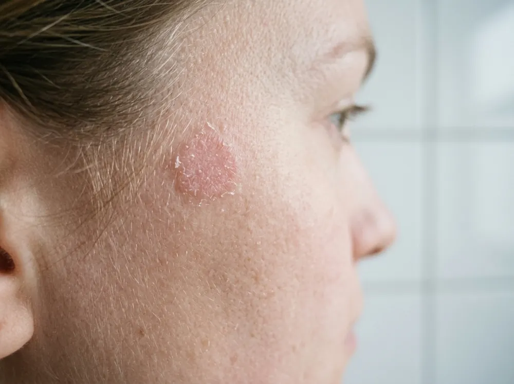
Pictures help, and they come with a caution about skin tone. DermNet states that in Māori, Pacific Islander and African patients the papular variant of atopic dermatitis is observed, that perifollicular and extensor patterns may be more common in patients of African descent, and that the redness can appear violaceous. Eczema pictures that show only pink patches on pale skin describe one version of the condition, and an eczema face on darker skin can look quite different.
Atopic, Contact or Seborrheic
Atopic
Atopic dermatitis is usually diagnosed clinically, according to DermNet, and investigations are not required. Patch testing should be considered, particularly if the dermatitis becomes resistant to treatment. Our article on eczema versus psoriasis covers the features that separate eczema from its main look-alike on the body.
Contact
Contact dermatitis is a reaction to something that touches the skin, either an irritant or an allergen. DermNet notes that the appearance of eyelid contact dermatitis is similar, whatever the cause. Our article on hand eczema covers how irritant, allergic and atopic types overlap on the hands, and the same overlap applies on the face.
Seborrheic
Seborrheic dermatitis is tied to sebum-rich skin. DermNet lists typical features: winter flares that improve in summer, minimal itch most of the time, combination oily and dry mid-facial skin, scaly red eyelid margins and salmon-pink, thin, scaly, ill-defined plaques in the skin folds on both sides of the face. On darker skin it may present as scaly, hypopigmented macules and patches, and DermNet says it is among the five most common diagnoses in Black patients. Children of colour often show redness, flaking and hypopigmentation of the affected areas and folds rather than classic cradle cap.
A fourth look-alike deserves a mention. Periocular dermatitis is a small-bump form of periorificial dermatitis, with small red scaly papules and pustules around the eyes, and DermNet says it often occurs in people using topical corticosteroids. Our article on perioral dermatitis covers the mouth version.
Eyelids Are Different
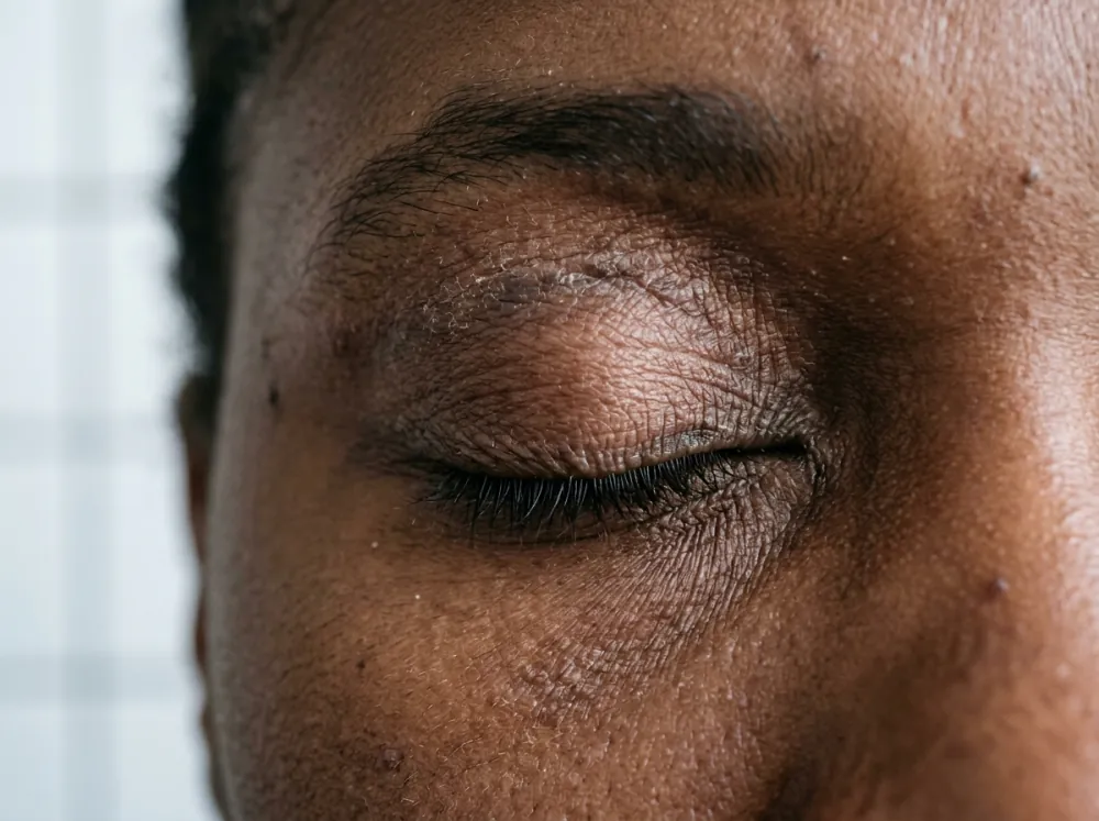
DermNet explains that the thin skin of the eyelids is particularly sensitive to irritants and allergens, and that contact with the same trigger may not cause a rash elsewhere. The lids may itch, sting or burn, appear red and scaly, and swell, and with persistence they thicken with increased skin markings (lichenification). The lid margins may become involved.
That sensitivity changes how the rash behaves. DermNet states that allergens can reach the eyelids indirectly, by transfer from the hands. It lists nail cosmetics, hair dye (paraphenylenediamine) and metals from coins or fastenings, and some airborne allergens such as dust mite and plant pollens, so an eyelid rash can come from something never applied to the eyelid.
The Figure Audited: "Facial Eczema Is Always Atopic"
The claim fails the data, and so does its mirror image, that it is always an allergy.
What the Patch-Test Clinics Found
A North American Contact Dermatitis Group analysis of 50,795 patients referred for patch testing between 1994 and 2016 found that 4.6% had eyelid dermatitis only and 3.2% had eyelid plus head or neck dermatitis (abstract). The final diagnoses were:
| Final diagnosis | Eyelid only | Eyelid plus head or neck |
|---|---|---|
| Allergic contact dermatitis | 43.4% | 53.5% |
| Irritant contact dermatitis | 17.0% | 9.8% |
| Atopic dermatitis | 13.1% | 13.8% |
The most frequent relevant allergens were nickel sulfate (18.6% and 22.5%), fragrance mix I (16.5% and 18.3%), methylisothiazolinone (16.5% and 17.7%), gold sodium thiosulfate (14.7% and 11.4%) and balsam of Peru (11.9% and 12.6%). Patients with eyelid involvement were more likely to be female, white and over 40, and to have a history of hay fever or atopic dermatitis.
A 2024 meta-analysis of 65 studies, with 21,793 patch-tested eyelid dermatitis patients, found atopic eyelid dermatitis in 27.5% (95% confidence interval 17.7 to 38.4), exceeding the previous estimate of 10%. Half of the 8,453 patients with reported distribution, 51.6%, had the eyelids affected in isolation.
Why the Numbers Disagree
The 13% and the 27.5% describe different samples and different definitions. Both come from people referred or tested for suspected allergy, who are not a random sample of everyone with facial eczema. Neither source says how many people never reach a patch-testing clinic. The meta-analysis also notes that the atopic eyelid share exceeded earlier estimates. As reasoning, not a finding, that suggests the figure depends on how studies counted it.
DermNet adds a further disagreement. It says allergic eyelid contact dermatitis is no more likely in people with atopic eczema or sensitive skin than in people without them. The clinic analysis found that eyelid patients were more likely to have a history of atopic dermatitis or hay fever, but it compared them with other patch-tested patients without eyelid involvement, not with the general population. The two statements are not directly comparable.
Verdict: the claim is false. In these clinic populations atopic dermatitis was a minority diagnosis for eyelid rashes, though not a rare one, and allergic and irritant contact dermatitis made up a larger share. The plan's idea that distribution tells you which type is also overstated: DermNet says eyelid contact dermatitis looks the same whatever the cause, and the clinic analysis lists the lack of specific distribution patterns as a limitation. Sorting the types usually needs history and often patch testing.
Triggers Worth Testing
Allergens
Patch testing is how allergens are identified. DermNet lists, for the eyelids, allergens in moisturisers, eye creams, sunscreens, makeup, cleansers and essential oils, nickel and gold in jewellery, nickel in eyelash curlers and tweezers, adhesives in false eyelashes, preservatives in eye drops and contact lens solution, and rubber in goggles and spectacle frames.
The male face has its own pattern. A North American analysis of 50,507 patients found that male facial dermatitis rose from 5.6% in 1994 to 1996 to 10.6% in 2015 to 2016 (abstract). Men with facial dermatitis were younger and commonly reacted to allergens in personal care products: preservatives, fragrances, hair dye and surfactants.
DermNet notes that allergic contact dermatitis usually appears one to several days after contact, which makes the cause hard to spot, and that allergy often develops after repeated previous contact, so the trigger is not necessarily something new.
Irritants
DermNet lists irritants for the eyelids: soaps and detergents, acids and alkalis, chlorine under swimming goggles, dust, drying agents and cosmetics such as eyeliner, eye shadow, mascara and sunscreen. Irritant dermatitis involves no allergy, and patch tests to suspected allergens are negative.
A trigger log of everything that touches the face, with dates, is the record a clinician can use. Whether in a notes app or a photo album or an app like Skinic, dated photographs taken in the same light make a flare comparable, and they show the clinician what the skin looked like when the appointment is days away.
Why Facial Skin Needs Gentler Handling
The same thin skin that makes the eyelids reactive shapes the advice. DermNet's general measures for eyelid contact dermatitis include avoiding contact with irritants and known allergens, avoiding rubbing and scratching, washing eyelids with plain water or a cream cleanser designed for sensitive skin, and avoiding eyelid cosmetics while the dermatitis is active. For anything detected by patch tests, avoidance is life-long.
On treatment, DermNet states that short courses of mild topical corticosteroids, such as hydrocortisone 1%, or calcineurin inhibitors may be needed to treat active inflammation, and that severe eyelid contact dermatitis is usually treated with a short course of oral corticosteroids. In seborrheic dermatitis it says calcineurin inhibitors are indicated if topical corticosteroids are needed frequently, because they have fewer adverse effects on facial skin with long-term use. This is reported guidance, and a clinician decides on it. Our articles on acne treatment that made redness worse and on skin barrier repair cover what steroids on the face can do and how a damaged barrier behaves.
When to Get Seen
A facial rash is a reason for a visit in these situations, drawn from the sources:
- An itchy eyelid rash that keeps coming back. Patch testing is described by the clinic analysis as a critical tool in evaluating eyelid dermatitis.
- A rash that stops responding. DermNet says patch testing should be considered in atopic dermatitis that becomes resistant to treatment.
- Eczema that may have become infected. DermNet lists eczema herpeticum and staphylococcal infection among the complications of atopic dermatitis, and what they look like is for a clinician to judge.
- Small red bumps and pustules around the eyes or mouth. That pattern may be periorificial dermatitis, not eczema. Our article on rosacea, perioral dermatitis and acne covers how those look-alikes differ.
- Eye symptoms, such as scaly, red lid margins. DermNet describes blepharitis in seborrheic and eyelid dermatitis.
- A first rash in adulthood, or one that is new, changing or unlike its neighbours.
Conclusion
Eczema on face and eyelid skin is a group of conditions that look alike. Atopic, allergic contact, irritant contact and seborrheic dermatitis share redness, scale and itch, and on darker skin the redness can read violet or brown, with colour change that lasts. In patch-test clinics, atopic dermatitis was a minority eyelid diagnosis (13% to 27.5% depending on the source), and allergic contact dermatitis was the largest.
Because the types look similar on the eyelids, history and patch testing do more than appearance. Keep a trigger log, photograph flares in the same light, and bring both to your own clinician.
Frequently asked questions
5 questions · tap one to open the answer
What does eczema on the face look like?
Is facial eczema always atopic?
What causes eczema on the eyelids?
How is seborrheic dermatitis different from eczema on the face?
When should I see someone about eczema on my face?
Medically reviewed by
Dr. M.M Hanaei
Aesthetic Physician, Dermatology Fellowship
- MD — Shahid Beheshti University of Medical Sciences (SBMU)
- MCC Licensure (Medical Council of Canada)
- Dermatology Fellowship, Switzerland
- Certified in advanced aesthetic procedures
Last reviewed 10 October 2026