Skinic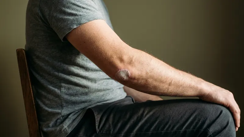
You have a patch. It has been there for weeks, it is scaly, and two people have given you two different answers about what it is.
Eczema and psoriasis are genuinely different diseases — different mechanisms, different treatments, different long-term implications — and they produce patches that can look strikingly similar, particularly early on and particularly to someone looking at their own arm in a bathroom.
The good news is that there are three or four features that separate them reliably, and none of them is the one most people focus on. Redness is the least useful signal here. The edge of the patch, where it sits on your body, and what your fingernails look like will all tell you more.
What This Page Will and Will Not Do
Before anything else, the scope.
This article will help you work out which of the two you are probably looking at, and what to say when you get an appointment. It will not tell you how to treat either one.
That is a deliberate limit, not a cop-out. Both conditions are chronic, immune-mediated diseases with genuine prescription treatment behind them, and the treatments diverge — what helps one can be irrelevant or unhelpful for the other. There is no over-the-counter route to managing either properly, and the most common way people lose years is by treating a confidently self-diagnosed condition that turns out to be the other one.
So: recognition here, treatment with a clinician. If you want the full differential for facial bumps rather than body patches, it might not be acne covers that separately.
Scale and Edge: The Two Things to Look At
If you take one thing from this article, take this: look at the border of the patch, not the middle of it.
Psoriasis: Silvery Scale, and a Border You Could Draw Around
Plaque psoriasis presents as red, scaly plaques with well-defined edges, and the scale is typically silvery white. Those two features together are close to a signature.
"Well-defined" is doing real work. A psoriasis plaque tends to stop. There is affected skin, there is normal skin, and the transition between them happens over a millimetre or two — you could trace the outline with a pen. The scale sits on top, thick and loosely adherent, and often looks lighter than the skin underneath it.
In skin folds, the presentation changes: plaques there appear shiny with a moist peeling surface rather than dry and silvery.
Eczema: A Patch That Fades Into Normal Skin
Atopic dermatitis does the opposite at the edge. There is rarely a line — the affected skin shades gradually into unaffected skin, and the outline is different depending on how hard you look.
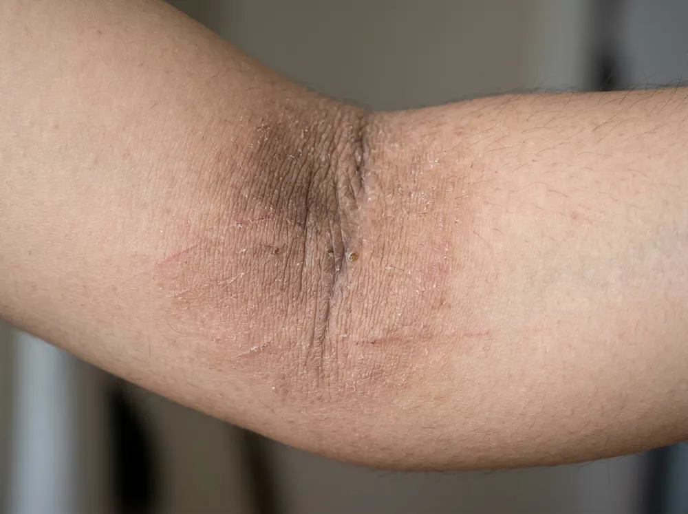
It also changes character over time. Acute eczema is red, weeping or crusted, and may have blisters. Later, the picture shifts: the skin becomes less red but thickened and scaly, a change called lichenification, and it may crack. That thickening is the consequence of months of scratching and rubbing, which is itself a clue.
So the quick read: a sharply bordered patch with silvery scale leans psoriasis; a vague-edged patch that has been weeping, or that has become leathery and thickened, leans eczema.
Where Each One Turns Up on the Body
Distribution is the second strong signal, and it is close to opposite between the two.
Psoriasis favours extensor surfaces — the outside of a joint. The most common sites are the scalp, elbows and knees, with chronic plaque psoriasis most often affecting elbows, knees and the lower back.
Eczema favours flexures — the inside of a joint. In toddlers and school-age children it becomes flexural, particularly involving the antecubital and popliteal fossae: the creases of the elbows and the backs of the knees. In infants it starts differently, on the scalp, in the armpits and groin creases, then the face. In adults it may stay flexural, become more diffuse, or settle on the hands, which has its own types and triggers.
| Psoriasis | Eczema | |
|---|---|---|
| Border | Well-defined, you could draw it | Fades into normal skin |
| Scale | Thick, silvery white | Fine, or crusting; later thickened and leathery |
| Favours | Outside of joints — elbows, knees, lower back, scalp | Inside of joints — elbow creases, backs of knees |
| Weeping | No | Yes, in acute phases |
| Nails | Pitting, onycholysis, yellowing, ridging | Usually unaffected |
| Itch | Mostly mild, sometimes severe | Intense — central to the condition |
| Age of onset | Peaks at 15–25 and 50–60 | Usually starts in infancy or childhood |
| Family pattern | About a third have affected relatives | Clusters with hay fever, asthma, food allergy |
Elbows and knees are the single most useful place to look, because the two conditions sit on opposite sides of the same joint.
Itch Is Not the Divider People Think It Is
A lot of comparison content says psoriasis does not itch. That is not accurate, and believing it sends people down the wrong path.
In psoriasis, itch is mostly mild but may be severe in some patients, leading to scratching and lichenification — the same thickening that is usually presented as an eczema feature. So severe itch does not rule psoriasis out.
What is true is that itch is central to atopic dermatitis in a way it is not for psoriasis. Eczema is usually described as generalised dryness, itch and rash, and the itch-scratch cycle is the engine of the disease — it is what converts a red patch into a leathery one.
The more useful question is not "does it itch" but "is the itch the worst part?" In eczema it very often is. In psoriasis the plaques themselves, and sometimes joint pain, tend to be what people lead with.
Check the Nails, and Ask About Relatives
Two pieces of evidence sit outside the patch entirely, and both are more reliable than staring at it.
Nails. Nail psoriasis causes pitting, onycholysis, yellowing and ridging — small dents in the nail plate, the nail lifting from its bed, discolouration. Eczema usually leaves nails alone. If you have a scaly patch on your elbow and pitted nails, that combination is meaningful, and it is worth mentioning specifically because nail involvement is associated with inflammatory arthritis, which changes what a clinician looks for.
Family and personal history. About a third of people with psoriasis have family members with psoriasis. Atopic dermatitis clusters differently — with hay fever, asthma and food allergies — and it clusters within the person as well as within the family. If you had eczema as a child, have hay fever now, and a relative has asthma, that atopic pattern is significant evidence.
Age helps too. Psoriasis onset peaks at 15–25 and again at 50–60. Atopic dermatitis usually begins in infancy or childhood, so a first-ever scaly patch appearing at 45 in someone with no history of childhood eczema is less likely to be it.
Why This Is Harder to See on Deeper Skin — and Matters More
Everything above leans on seeing colour and contrast, and that is where visual diagnosis breaks down.
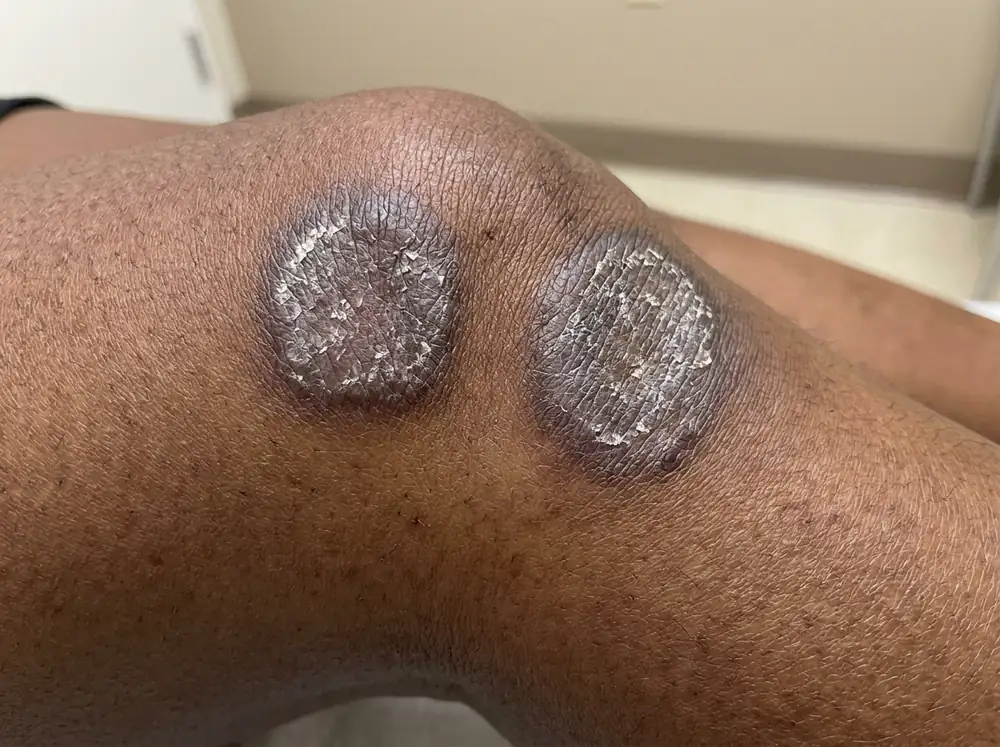
Erythema is redness from increased blood flow, and in richly pigmented skin melanin masks it. Rather than reading as red, inflammation in Fitzpatrick types IV to VI may present as violaceous, grey or brown hues. The documented consequences are serious: both atopic dermatitis and psoriasis go unrecognised when the expected redness is not visible, and severity is systematically underestimated because the standard scoring systems are built around erythema. The same paper reports that Black children are nearly six times more likely to present with severe disease.
The presentations differ in other ways too. In darker skin, psoriasis plaques are typically thicker with a more pronounced silver scale and more itch, and thick plaques may look violet or dark rather than red. And in both conditions, clearance frequently leaves postinflammatory pigment change behind — which in atopic dermatitis is described as more noticeable than in white skin and a source of significant psychological distress. Often the pigment outlasts the disease and becomes the thing people actually want treated; hyperpigmentation in brown and deep skin tones covers that.
Two practical adjustments follow. Go by the non-colour signs — the border, the scale, the distribution, the nails, the thickening. And when describing symptoms, name the inflammatory signs rather than the colour: swelling, warmth, pain, tightness. That shift is exactly what the literature recommends, and it is more accurate for everyone.
If you are unsure of your own phototype, Fitzpatrick skin types I–VI sets out how the scale works and what it does and does not predict.
When It Is Both
One honest complication, because a clean binary is not quite the truth.
The two conditions overlap more than the textbook split suggests. They share immune pathways — IL-17 and IL-22 appear in both — and 81% of gene disruptions found in atopic dermatitis also occur in psoriasis. Among children with psoriasis, reported coexistence with atopic dermatitis ranges from 3.7% to 25.9% depending on the study, though the authors note that co-occurrence in the same individual is less frequent than might be expected, and some researchers now argue for treating overlapping cases as their own category.
What that means for you is modest but worth knowing: if your patches do not fit neatly into either column, that is a recognised situation rather than a failure of observation — and it is a particularly good reason to stop trying to settle it yourself.
What to Bring to the Appointment
Diagnosis here is clinical, which means it rests on what a clinician sees and what you tell them. Three things make that appointment substantially better.
Dated photographs across a flare. Both conditions fluctuate, and the day you get an appointment is rarely the day your skin looks worst. Photograph in consistent conditions — same spot, same distance, same daylight, no filters — and include the edge of a patch in close-up, since the border is the most diagnostic feature you can capture. Whether that lives in a dated album on your phone or in something built for it like Skinic, a clear photograph from a bad week is real clinical information.
A timeline and a family history. When it started, whether you had eczema as a child, whether you have hay fever or asthma, and whether anyone related to you has psoriasis, eczema or psoriatic arthritis.
Your hands, and anything else affected. Show your nails without polish. Mention the scalp, the ears, the navel, the natal cleft and any joint pain, even if you think they are unrelated — those are exactly the sites that settle the question, and people routinely present one patch while leaving the informative ones covered.
Conclusion
Stop looking at the redness. It is the least reliable feature of the two conditions, and on deeper skin tones it may not be visible at all.
Look at the edge. A patch with a border you could trace, carrying thick silvery scale, on the outside of an elbow or knee, in someone with pitted nails and a relative who has it too — that is a psoriasis pattern. A patch with no clear border that has wept or crusted, sitting in the crease of an elbow or behind a knee, in someone who itches constantly and has hay fever — that is an eczema pattern.
Then stop. Both are chronic immune-mediated diseases, both have real treatment, and both are diagnosed by a clinician rather than by an article. What this page can do is make sure you arrive at that appointment with the right photographs, the right history, and your nails visible.
That is a genuinely better outcome than another month of guessing — and a much better one than a year of treating the wrong disease.
Frequently asked questions
5 questions · tap one to open the answer
How can you tell eczema and psoriasis apart?
Does psoriasis itch?
Can you have eczema and psoriasis at the same time?
Why is it harder to diagnose eczema or psoriasis on darker skin?
Do I need to see a doctor for eczema or psoriasis?
Medically reviewed by
Dr. M.M Hanaei
Aesthetic Physician, Dermatology Fellowship
- MD — Shahid Beheshti University of Medical Sciences (SBMU)
- MCC Licensure (Medical Council of Canada)
- Dermatology Fellowship, Switzerland
- Certified in advanced aesthetic procedures
Last reviewed 14 September 2026