Skinic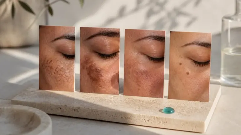
You have probably stood in a skincare aisle, stared at a shelf full of dark spot correctors, and picked one based on the prettiest packaging or the boldest claim. Most people do. And most people end up disappointed, not because the product was low quality, but because it was designed for a completely different type of dark spot than the one on their face.
Dark spots on face are not a single condition. The term covers at least three distinct types of hyperpigmentation, each with its own cause, behavior, and treatment logic. Melasma, sun spots, and post-inflammatory hyperpigmentation look similar on the surface but respond to completely different approaches. Treating one like another does not just waste money; it can actively make the problem worse.
This guide will walk you through everything you need to know as a beginner: how to identify which type of dark spot you are dealing with, which ingredients are backed by evidence for each type, when topical products are not enough, and the common mistakes that keep dark spots from fading. Start with the right information, and the right treatment becomes far more obvious.
Why Most Dark Spot Treatments Fail Before You Even Open the Bottle
Pick up any "dark spot serum" at a pharmacy and the label will promise to work on all types of hyperpigmentation. That promise is, at best, half true.
Dark spots on the face are not a single condition. Melasma, solar lentigines, and post-inflammatory hyperpigmentation (PIH) each arise through distinct biological mechanisms, sit at different depths within the skin, and respond to entirely different treatment strategies. An ingredient clinically proven to fade one type can be ineffective, or actively counterproductive, against another.
This is why products fail. Not because they are low quality, but because the active ingredient was matched to the wrong mechanism. Research confirms that even within a single type like PIH, the depth of melanin deposit determines whether a topical treatment can reach the pigment at all. A serum working at the epidermal level does nothing for pigment lodged in the dermis.
The hyperpigmentation market compounds this problem by selling "universal" formulas targeting one pathway while packaging them as solutions for every dark spot. Consumers follow the instructions, see no results after weeks, and conclude the ingredient does not work, when the real issue was misidentification from the start.
Dermatologists do not skip this step. Correct lesion identification is established clinical practice before any treatment direction is recommended, because skin type, pigmentation depth, and underlying trigger all determine what will actually work.
This guide covers exactly that process: what each type looks like, what causes it, which ingredients and procedures are matched to each, and what commonly makes each one worse.
The Three Main Types of Dark Spots on the Face
Each of the three types operates through a different biological mechanism, which is precisely why a serum that works on one can be ineffective or even counterproductive on another.
Melasma
Melasma appears as large, blotchy, symmetrical patches with irregular borders that blend gradually into surrounding skin rather than stopping at a defined edge. It concentrates on the cheeks, forehead, upper lip, and bridge of the nose. The driving forces are hormonal fluctuations combined with UV exposure, and the condition involves multiple systemic factors: skin barrier disruption, a vascular response, chronic low-grade inflammation, and pigmentation that extends into the deeper dermal layers. This biological complexity is what makes melasma the most difficult of the three to treat.
Solar Lentigines (Sun Spots or Age Spots)
Solar lentigines are small, well-defined brown spots with relatively sharp, visible borders. They develop on areas with the highest cumulative UV exposure: cheeks, nose, and the backs of hands. Unlike melasma, they are concentrated in the shallow epidermal layers of skin, caused by localised overproduction of melanin in clusters. That epidermal location makes them more accessible to both topical ingredients and targeted procedures.
Post-Inflammatory Hyperpigmentation (PIH)
PIH develops after skin inflammation or injury, including acne, rashes, burns, scratches, or any dermatological trauma. The result is tan to brown flat marks that remain after the original inflammation resolves. PIH is not a standalone pigmentation condition; it is the skin's melanin response to injury. Depth varies with the severity of the original inflammation, which directly affects how long marks persist and how they respond to treatment.
All three can appear simultaneously on the same face. That overlap is what makes self-diagnosis genuinely difficult, and it is why identifying each type before selecting a treatment is the only approach that avoids wasted effort and spending.
Melasma: The Hormonal Dark Spot That Requires a Different Strategy
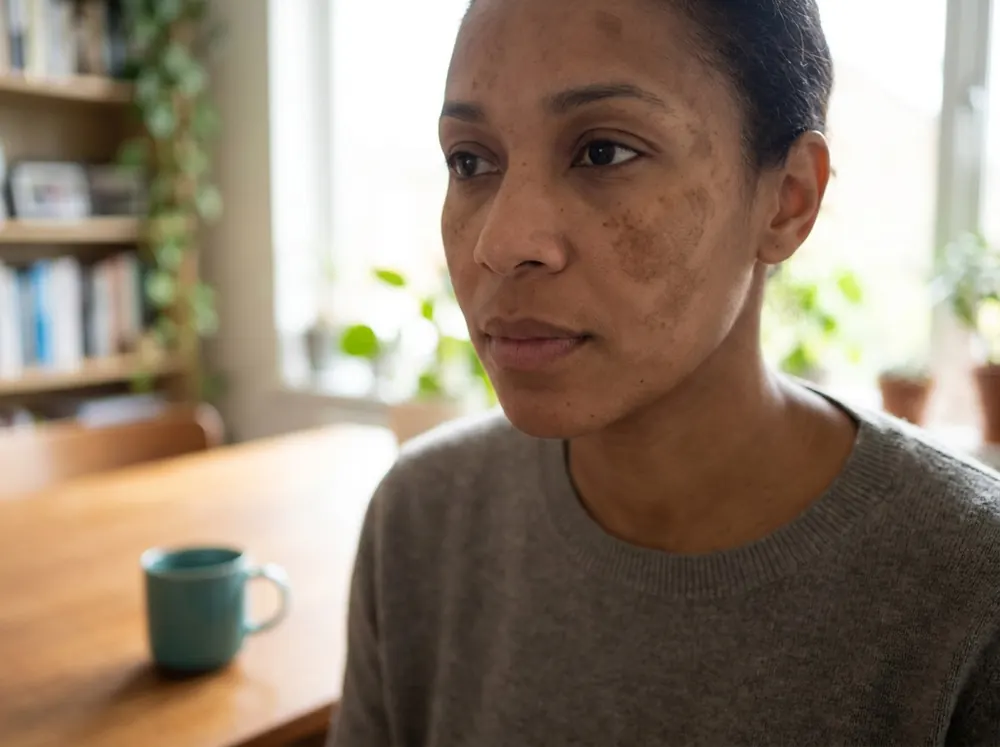
Of the three dark spot types, melasma is the most frequently mismanaged, and the consequences of getting it wrong can actively set your skin back.
Who gets melasma follows a clear pattern. It is significantly more common in women and disproportionately affects people with darker skin tones, particularly Fitzpatrick types III to VI. This skin-tone distribution matters because treatment approaches that suit lighter skin can cause additional pigmentation damage in deeper tones. If you do not know your own type, how to find your Fitzpatrick skin type works through the two questions that place you.
Hormonal triggers are the defining feature. Pregnancy is so commonly associated with melasma that the condition carries the nickname "mask of pregnancy," with up to 50% of pregnant women developing it. Oral contraceptives, hormone replacement therapy, and thyroid dysfunction are equally established triggers. This is why topical treatment alone often underperforms: if the hormonal driver is still active, the melanocytes continue receiving the signal to overproduce pigment.
UV exposure is the other half of the equation. Melasma is not caused by sun exposure alone, but UV acts as a co-trigger that amplifies hormonal stimulation. Even low-level exposure, such as incidental daylight through a window, is sufficient to activate a flare. Sun protection is not a supporting measure for melasma; it is foundational, and no topical ingredient performs well without it.
The mechanism is the reason standard spot treatments fail. Unlike solar lentigines, which sit in the superficial epidermis, melasma involves deeper skin layers, the vascular network, and a broad inflammatory response. Applying a concentrated brightening serum to individual patches, as most over-the-counter products are designed to do, addresses the wrong target. The correct approach is managing the broader affected area to control an overall background colour, not erasing spots one by one. Aggressive localised treatment can irritate the skin and trigger further melanin production, worsening the very problem you are trying to solve.
Finally, recurrence is the rule rather than the exception. Successfully fading melasma does not mean it is resolved. Without ongoing UV protection and attention to hormonal triggers, it reliably returns.
Sun Spots and Age Spots: The Most Treatable Type of Hyperpigmentation
Unlike melasma, solar lentigines sit squarely in the superficial epidermal layers of skin. That anatomical fact is what makes them the most straightforward type of hyperpigmentation to treat. Topical ingredients reach epidermal melanin relatively easily, and targeted procedures can address individual spots without disturbing the broader skin surface.
Their other defining feature reinforces this advantage: well-defined, clearly demarcated borders that separate each spot from surrounding skin. This makes selective spot treatment both logical and effective. With melasma, as covered above, that same approach backfires because the condition has no clean boundary and involves systemic mechanisms beneath the surface.
What causes them is accumulated UV exposure over years and decades, not a single sunburn or hormonal shift. Melanin clusters form where the skin has absorbed the most lifetime ultraviolet radiation, which is why solar lentigines concentrate on the face, backs of hands, and forearms, and why they become increasingly common after middle age. Up to 90% of Caucasians over 60 have at least some solar lentigines, reflecting cumulative photodamage rather than any underlying systemic condition.
For procedural treatment, 532 nm pigment lasers are a common clinical choice. Picosecond lasers are increasingly compared with traditional nanosecond Q-switched lasers for this indication; they use shorter pulses to fragment pigment through a photomechanical effect rather than thermal energy, with the theoretical benefit of reducing heat spread to surrounding tissue. Results from procedures are typically faster and more dramatic than topical alternatives.
Topical options with epidermal melanin-inhibiting activity, including vitamin C, alpha-arbutin, kojic acid, and retinoids, can produce meaningful fading with consistent use. Results take longer than procedural treatment, measured in months rather than sessions, but the cost barrier is considerably lower.
Prevention, however, is the most cost-effective strategy of all. New solar lentigines will continue forming with ongoing UV exposure regardless of how many existing spots are treated. Broad-spectrum SPF applied daily is not optional maintenance; it is the mechanism that stops treatment from running on a treadmill.
Post-Inflammatory Hyperpigmentation: When Your Skin's Healing Response Leaves a Mark
Unlike sun spots, which result from accumulated UV exposure, post-inflammatory hyperpigmentation is not a pigmentation condition in its own right. It is the skin's melanin response to injury or inflammation. Acne, rashes, burns, scratches, and even some skincare procedures can all trigger it. This distinction matters for treatment: managing the source of inflammation, such as active acne, is part of the plan, not a separate concern.
PIH marks are flat, tan to dark brown, and appear precisely where prior inflammation occurred. They are sometimes mistaken for scars, but the difference is structural. Atrophic (depressed) or raised scarring involves physical changes to dermal tissue. PIH is pigment only, with no change in skin texture or surface.
How long PIH takes to fade depends heavily on skin tone. In lighter skin tones, marks may resolve on their own within 3 to 6 months. In deeper skin tones, the same marks can persist for a year or longer without treatment, because higher baseline melanin activity amplifies the post-inflammatory response. This is also why PIH is among the most common reasons people with darker skin tones seek a dermatologist.
Depth adds another variable. Superficial PIH confined to the epidermis responds well to topical treatment. PIH that extends into the dermis, typically from more severe or prolonged inflammation, is more resistant and may require professional procedural support to shift.
The single most avoidable worsening factor is picking. Squeezing a pimple or scratching inflamed skin creates an additional trauma event, triggering a fresh inflammatory response and producing more melanin. The resulting mark is often darker and longer-lasting than it would have been otherwise.
Because the underlying mechanism is inflammation driving excess melanin production, the most effective topical approach combines anti-inflammatory ingredients with melanin-suppressing actives. Addressing one without the other leaves part of the process unchecked.
How to Tell Your Dark Spots Apart: A Practical Identification Guide
Knowing what each type looks like is the practical bridge between understanding these conditions and choosing a treatment that actually works. Four markers will get you most of the way there.
Shape and border. Melasma presents as irregular, blotchy patches that blend gradually into the surrounding skin with no clear edge. Solar lentigines have well-defined, relatively sharp borders; the boundary between spot and skin is visible. PIH is flat and confined precisely to the site of prior inflammation, matching the footprint of the original blemish or wound.
Symmetry. This is the most reliable single clue for melasma. It almost always appears on both sides of the face in a mirrored pattern, because its hormonal driver affects the whole face rather than a specific location. Solar lentigines and PIH follow no symmetrical pattern; they appear where UV exposure or inflammation happened to occur.
Trigger history. Think back to when the spots first appeared or worsened. Did they emerge during pregnancy, while taking hormonal contraceptives, or after a period of heavy sun exposure? That points strongly to melasma. Spots that have gradually accumulated over years on sun-exposed skin suggest solar lentigines. If every mark can be traced to a specific healed blemish, rash, or skin injury, that is PIH.
Location. Melasma concentrates on the cheeks, forehead, upper lip, and nose bridge. Solar lentigines cluster on the highest UV-exposure zones, primarily the cheeks and nose. PIH has no preferred location; it appears wherever inflammation previously occurred.
Skin tone. Both melasma and PIH are more prevalent and more persistent in deeper skin tones, which is clinically significant because some treatments suitable for fairer skin can worsen pigmentation in darker complexions. Solar lentigines occur across all skin tones with sufficient UV exposure history.
When your spots do not fit cleanly into one category, or when more than one type appears to coexist, committing to a treatment before confirming the type is a reliable way to waste money. Price is not the shortcut either — what "medical grade" skincare actually means sets out which product distinctions are real and which are only on the label. A structured skin analysis or dermatologist assessment provides the clarity needed to match your approach to what you actually have.
Which Ingredients Actually Work for Each Type of Hyperpigmentation
Once you've identified your spot type, ingredient selection becomes straightforward: match the active to the mechanism.
Melasma
Because melasma involves vascular, inflammatory, and hormonal pathways, standard tyrosinase inhibitors alone often fall short.
- Tranexamic acid targets the vascular and inflammatory components that other brightening ingredients miss. Meta-analysis data shows it produces clinically meaningful reductions in Melasma Area and Severity Index scores, and recurrence rates are significantly lower when it is included alongside other treatments.
- Niacinamide blocks melanosome transfer to skin cells and supports barrier function, addressing two of melasma's several mechanisms in one ingredient.
- Azelaic acid is well-tolerated across skin tones and delivers both pigmentation inhibition and anti-inflammatory activity.
- Hydroquinone 2–4% remains a clinical benchmark but requires supervised use and treatment cycling to prevent rebound darkening.
Solar Lentigines
Epidermal pigmentation responds well to tyrosinase inhibitors and cell-turnover actives.
- Vitamin C (L-ascorbic acid) inhibits tyrosinase and provides antioxidant protection against ongoing UV-triggered melanin production.
- Alpha-arbutin and kojic acid are effective tyrosinase inhibitors well-suited to epidermal pigmentation. The trials behind kojic acid used leave-on creams over 12 to 16 weeks.
- Retinoids accelerate cell turnover, shedding pigmented surface cells and improving penetration of other actives.
Post-Inflammatory Hyperpigmentation
Treating the PIH mark and controlling the inflammatory source are equally necessary steps.
- Niacinamide is among the most evidence-supported options, calming residual inflammation while reducing melanin transfer.
- Azelaic acid covers both anti-inflammatory and pigmentation-suppressing activity in a single ingredient.
- Retinoids reduce PIH depth over time by accelerating epidermal turnover.
- Addressing the underlying trigger, such as active acne, is not optional; uncontrolled inflammation will continue producing new marks.
Universal Rule and Cautions
Broad-spectrum SPF 30 or higher is the one intervention that applies to all three types. UV exposure continuously restimulates melanin production, so any topical treatment is undermined without it applied daily.
Use these ingredients carefully without professional guidance: high-strength AHAs and physical exfoliants create micro-inflammation that worsens PIH and can trigger melasma flares. Vitamin C concentrations above 20% risk irritating reactive skin tones and paradoxically deepening PIH.
When Topicals Are Not Enough: Procedural Treatments by Spot Type
When consistent topical use has not delivered adequate results, procedural options become relevant, but the right procedure depends entirely on which type of hyperpigmentation you have.
Solar lentigines are the strongest candidates for procedural treatment. Their epidermal location and well-defined borders make them accessible to targeted energy-based devices. 532 nm KTP and Nd:YAG lasers are among the most commonly used. Picosecond lasers offer a theoretical advantage over traditional nanosecond Q-switched devices: they fragment pigment through a photomechanical effect rather than thermal energy, potentially reducing collateral skin damage. Clinical data supports this distinction, picosecond devices have demonstrated 67.9% to 93.02% clearance rates for solar lentigines, compared to 36.36% to 76.6% for Q-switched nanosecond lasers.
Melasma requires a fundamentally different approach. Laser and IPL treatments that are appropriate for solar lentigines can trigger a post-inflammatory melasma flare, because the thermal response activates the same inflammatory pathway driving the condition. Any procedural melasma treatment should be preceded by topical priming and performed by a provider with specific melasma experience, not general pigmentation experience.
PIH may respond to superficial chemical peels using glycolic or lactic acid for epidermal cases. Deeper PIH and PIH in darker skin tones requires careful selection of peel depth; going too deep risks triggering additional post-inflammatory response and worsening the mark.
Fractional laser resurfacing is sometimes used for deeper PIH and topical-resistant melasma. In darker skin tones, fractional devices must be calibrated to minimise thermal spread and post-treatment inflammation.
On cost: topical regimens are significantly more accessible than procedural treatments. For solar lentigines, procedures can deliver faster and more dramatic results. For melasma, however, topicals combined with strict sun protection frequently outperform aggressive procedures over the long term, because procedures carry a real recurrence risk.
Common Mistakes That Make Dark Spots on Your Face Worse
Even the right treatment can backfire if your daily habits are working against it. These five mistakes are among the most common reasons hyperpigmentation stalls or worsens.
Spot-treating melasma like a sun spot. Because melasma is driven by hormonal signals and a broader inflammatory response across the skin, dabbing a high-potency brightening serum onto individual patches misses the mechanism entirely. Concentrated application can also irritate the skin, and that irritation triggers more melanin production, making the condition worse rather than better. Melasma requires treating the whole affected area, not individual marks.
Skipping or inconsistently applying SPF. UV exposure is an active co-trigger for melasma and solar lentigines, and a direct worsening factor for PIH. UVA rays penetrate window glass, so proximity to windows indoors carries real risk. Every topical treatment you apply is partially undermined each time SPF is missed, because the skin is simultaneously being stimulated to produce more melanin.
Over-exfoliating to accelerate fading. Excessive use of AHAs, physical scrubs, or retinoids creates micro-inflammation in the skin. That inflammatory signal drives the same melanin overproduction pathway that causes PIH in the first place, and it can trigger melasma flares. More exfoliation does not mean faster results; it often means slower ones.
Picking or squeezing blemishes. Each time you pick at a healing pimple, you convert it into a new trauma event. The additional inflammation extends the PIH response and frequently produces a darker, longer-lasting mark than if the blemish had healed undisturbed.
Cycling through products too quickly. Topical hyperpigmentation treatments generally require eight to twelve weeks of consistent use before visible fading occurs. Switching products every few weeks makes it impossible to determine what is actually working.
Prevention and Long-Term Maintenance After Treatment
Avoiding common mistakes protects your progress, but preventing recurrence requires building habits that actively hold results in place.
Daily broad-spectrum SPF 30 or higher is the single most supported preventive measure across all three dark spot types. Apply it every morning as the final step of your routine, and reapply every two hours during outdoor activity. If your skin is sensitive or reactive, look for physical sunscreens containing zinc oxide or titanium dioxide; these mineral filters provide reliable broad-spectrum coverage with fewer irritation concerns than some chemical alternatives.
For melasma, sun protection alone is not always sufficient. If the hormonal trigger driving your melasma, such as a specific oral contraceptive or hormone replacement therapy, remains in place, pigmentation will continue returning regardless of how consistently you treat it topically. Addressing the underlying hormonal cause is part of a complete management plan; a conversation with your GP or specialist about your options is worth having alongside any topical routine.
Adding an antioxidant serum to your morning routine provides a layer of protection that sunscreen cannot fully replicate. Vitamin C and niacinamide help neutralise UV-induced free radical damage and suppress the melanin stimulation that drives ongoing pigmentation. Apply your antioxidant serum before sunscreen for best effect.
Once your pigmentation has faded, do not stop entirely. Gradually reducing, rather than abandoning, your active ingredients reduces the risk of gradual recurrence, particularly as UV levels increase through spring and summer.
Finally, physical sun avoidance compounds everything else. Wide-brim hats and shade-seeking during peak UV hours (10am to 4pm) meaningfully reduce your cumulative exposure. For melasma patients this matters especially, as the condition can respond to UV levels too low to produce any visible redness.
When to See a Dermatologist About Hyperpigmentation
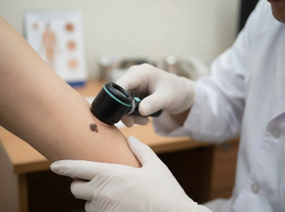
Preventive habits reduce recurrence, but they do not resolve every case. Knowing when to stop self-managing and seek professional input is an important part of any hyperpigmentation plan.
Seek evaluation promptly if a dark spot appears suddenly, grows rapidly, has irregular or multicoloured borders, or shows texture changes such as raised, rough, or scaly patches. These features can overlap with skin conditions requiring medical assessment and should not be managed with over-the-counter products while waiting to see whether they change.
For melasma, if consistent topical treatment over three to four months produces no visible improvement, or if spots continue darkening despite diligent sun protection, professional guidance is warranted. Prescription-strength formulations such as higher-concentration hydroquinone and combination therapy protocols require physician oversight and cannot be replicated with retail products.
For PIH, marks persisting beyond 12 months without meaningful fading, particularly in deeper skin tones, may indicate dermal pigmentation rather than epidermal, or an unresolved underlying inflammatory condition. A dermatologist can determine which is the case and direct treatment accordingly.
Dermatologists have access to diagnostic tools that change treatment decisions significantly. Dermoscopy allows visualisation of pigment at the dermal level, and a Wood's lamp examination can differentiate epidermal from dermal pigmentation. That distinction alone determines whether aggressive topical treatment is likely to work or whether a procedural approach is necessary.
If you are preparing for a dermatologist appointment, Skinic's skin analysis can help you document visible pigmentation patterns, organise your concerns, and generate a shareable report to bring with you. It bridges the gap between self-observation and clinical review without replacing the assessment a dermatologist provides.
Identify First, Treat Second: The Right Approach to Fading Dark Spots
Every dark spot on your face has a mechanism behind it. Identifying that mechanism is what separates a treatment plan that works from one that simply costs money.
The three types covered in this guide operate differently. Melasma is hormonal and systemic, requiring ingredients that address vascular and inflammatory pathways, such as tranexamic acid and azelaic acid. Solar lentigines are epidermal and UV-driven, responding well to tyrosinase inhibitors like vitamin C and cell-turnover accelerators like retinoids. PIH is an inflammation-triggered melanin response; managing the original inflammatory cause alongside niacinamide and azelaic acid gives it the best chance of resolving.
One factor applies regardless of type: daily broad-spectrum SPF. UV exposure actively restimulates melanin production in all three conditions. Without it, every other active ingredient is working against a current that never switches off. Sunscreen is not a finishing step; it is the foundation that makes every other treatment more effective.
If you have used a targeted product consistently for 8 to 12 weeks and seen no improvement, the issue is rarely the product's potency. Clinical evidence uses 12 weeks as the standard window to evaluate treatment efficacy, and falling short of that often means assessment happens too early. When it does not work within that window, the more likely explanation is a mismatch between the product's mechanism and your specific type of hyperpigmentation.
Before spending further, clarify what you are actually treating. A structured skin analysis or dermatologist consultation gives you that clarity. The right active ingredient, matched to the right condition, applied consistently under daily SPF protection, is a straightforward framework. Identification is simply the step that makes everything else count.
Conclusion
Dark spots are not a single problem, and they do not respond to a single solution. The type you have determines the treatment that works. Melasma requires hormone-aware, inflammation-minimizing care. Sun spots respond well to tyrosinase inhibitors and cell turnover support. PIH clears fastest when the inflammatory trigger is addressed alongside targeted actives.
Across all three, daily broad-spectrum SPF is non-negotiable. Without it, no active ingredient can deliver its full potential.
If previous treatments have disappointed you, the problem is likely identification, not effort. Start there. Examine your spots carefully, use the guidance in this post, and consult a dermatologist if you need confirmation.
The right ingredient, matched to the right condition, applied consistently under SPF protection, changes everything. Identify first, treat second, and give your skin the time it needs to respond.
Frequently asked questions
5 questions · tap one to open the answer
How can I tell if my dark spots are melasma, sun spots, or PIH?
Why isn't my dark spot serum working?
What ingredients actually work for melasma versus sun spots?
Can laser treatment make melasma worse?
Why do dark marks take longer to fade in darker skin tones?
Medically reviewed by
Dr. M.M Hanaei
Aesthetic Physician, Dermatology Fellowship
- MD — Shahid Beheshti University of Medical Sciences (SBMU)
- MCC Licensure (Medical Council of Canada)
- Dermatology Fellowship, Switzerland
- Certified in advanced aesthetic procedures
Last reviewed 6 September 2026