Skinic
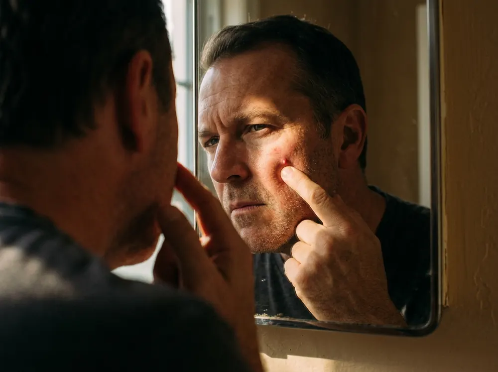
There is a particular bump that defeats everyone. Small, soft, slightly yellow, sitting on a forehead or a cheek. It looks exactly like something that should come out. You press it, and nothing happens. You press harder, and still nothing happens, and now it is red for two days.
That is not a failure of technique. There is nothing inside to extract.
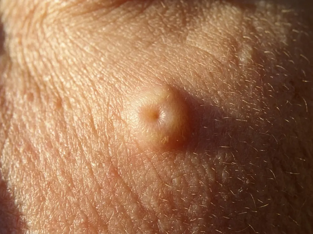
The lesion is not a pore with something stuck in it. It is a sebaceous gland that has enlarged, and the little crater in the middle — the thing that looks so much like a blackhead's opening — is the duct the gland drains through. It is an exit, not a plug.
Which explains the other thing people notice: it does not respond to anything aimed at oily skin, and it tends to arrive in middle age rather than in the years when skin is actually oily. The reason for that is the most counterintuitive part of this, and it is worth getting to properly.
Nothing Comes Out, Because There Is Nothing In There
Start with what the tissue actually is, because the histology settles the question completely.
When one of these is biopsied, what comes back is a dome-shaped lesion with a central dilated infundibulum attached to multiple lobules of sebaceous glands. In plain terms: a widened duct opening with a cluster of enlarged gland lobules hanging off it.
Gland tissue. Not retained keratin, not hardened sebum, not a plug of anything. A comedone has contents — that is what a comedone is. This has structure.
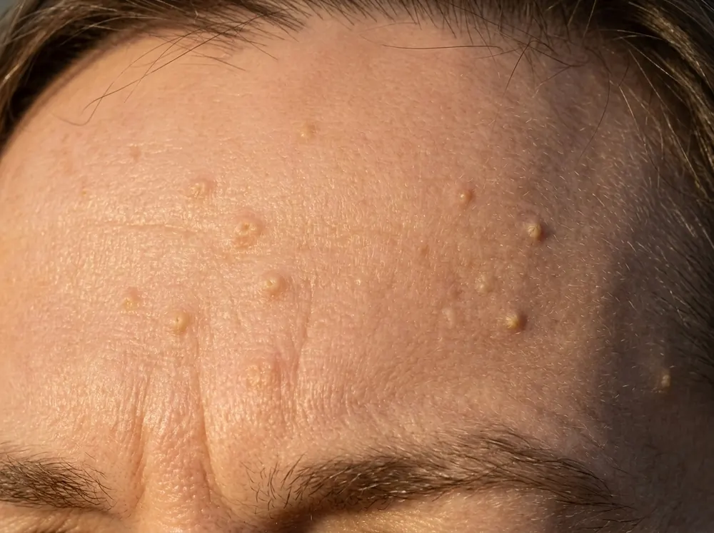
DermNet describes it as enlarged sebaceous glands on the forehead or cheeks of middle-aged and older people, and classifies it as a form of benign hair follicle tumour. The appearance is "small yellow bumps up to 3 mm in diameter" with "a central hair follicle surrounded by yellowish lobules."
So the squeezing question has a definitive answer. There is no version of pressing one of these that produces anything, because the only way to remove the lesion is to remove the gland — and the clinical literature is explicit that the entire sebaceous unit must be eliminated to prevent a recurrence. Partial destruction regrows.
What squeezing does achieve is inflammation around a structure that was not inflamed, in an area of the face where post-inflammatory marks are slow to fade.
Why It Arrives With Age, and Why That Is Backwards
Here is the part that makes everything else make sense.
Sebaceous glands are androgen-driven. Sebocytes carry the enzymes that convert weaker circulating androgens into dihydrotestosterone, which is what drives gland activity. That is the acne story: more androgen, more sebum, more trouble.
Sebaceous hyperplasia is the opposite. Declining androgen levels lead to a decrease in sebocyte turnover, which activates a feedback stimulation of sebocyte proliferation.
Follow the mechanism, because it is elegant. A sebocyte's normal life cycle — produced at the base, filling with lipid, migrating inward, disintegrating to release sebum — takes approximately one month. As androgens fall with age, the cells become smaller, carry less lipid, and migrate more slowly. They are produced at the usual rate and cleared more slowly than they arrive.
So they accumulate. The gland fills with cells that have not yet broken down and gets physically larger, and the duct widens with it.
The bump is a traffic jam, not a blockage. Which is why it appears in men typically around the eighth decade and in women shortly after menopause — both points where androgen levels drop. And why nothing that targets oily skin does anything: your skin at that age is producing less sebum, not more. If you have been treating these with products aimed at congestion, that is why none of it worked.
Who Gets Them
Roughly 1% of the healthy population, which understates how commonly people notice them, because the figure counts diagnosed cases rather than bumps on faces.
Two groups sit well outside that. Neonates — one prospective study of 1,000 consecutive newborns found sebaceous hyperplasia in 43.7% of them, driven by maternal hormones and resolving on its own within weeks.
And people on long-term immunosuppression. The association with ciclosporin is well established and the figures vary: StatPearls reports prevalence "as high as 10 to 16%" with long-term ciclosporin A, while a case report in a dermatology journal puts ciclosporin-induced sebaceous hyperplasia at "up to 30% of renal transplant patients." That is a wide spread, and picking a number between them would be inventing precision — the honest statement is that it is common enough in that population to be expected rather than investigated.
Systemic corticosteroids can do it too, and fast. The same report describes a 46-year-old man who developed multiple 1 to 3 mm umbilicated papules on his forehead and cheeks two weeks after starting prednisone at 40 mg/day.
The Central Dell, and the Three Things It Gets Mistaken For
The defining feature is the dimple. Lesions run 2 to 9 mm, skin-coloured or yellowish, with a central dell corresponding to the follicular infundibular ostium — the gland's drainage opening.
Here is how it separates from the things it resembles.
| Sebaceous hyperplasia | Milia | Closed comedones | |
|---|---|---|---|
| Colour | Yellowish or skin-coloured | White, distinctly | Skin-coloured to white |
| Centre | Depressed — a central dell | Domed, no dimple | Domed or a tiny opening |
| Contents | None; it is gland tissue | A keratin cyst | Keratin and sebum |
| Typical age | Middle age onward | Any age | Teens to thirties |
| Responds to extraction | No | Yes, with a lancet | Yes |
The site covers milia and closed comedones in their own right, and the full differential for every bump on the face sits alongside both. The short version: yellow plus a dimple plus middle age points one way, and nothing coming out confirms it.
The One That Matters: Basal Cell Carcinoma
There is a fourth possibility and it is the reason this article has a referral section rather than a reassurance.
Basal cell carcinoma can look like this. DermNet notes plainly that the lesions "are sometimes confused with basal cell carcinoma." StatPearls goes further: in a solitary facial lesion, a biopsy may be necessary to rule out basal cell carcinoma.
Both are small, both can be pearly or translucent, both occur on sun-exposed facial skin in older adults, and both have visible blood vessels. That is a lot of overlap on a face that has had decades of sun.
Dermoscopy distinguishes them, and the distinction is genuinely elegant — but it requires a dermatoscope and someone trained to read one, which is the point rather than a caveat. The two vascular patterns are described precisely:
Crown vessels, which point to sebaceous hyperplasia, are "long, out-of-focus telangiectasias, with tortuous yet regular barely branching vessels surrounding a central polylobulated whitish-yellowish area" — and critically, "they characteristically do not cross the center of the lesion."
Arborizing vessels, which point to basal cell carcinoma, are "large-diameter vessels that branch at irregular intervals into increasingly thin capillaries; they are bright red, have abrupt border cutoffs, and are in sharp focus." They do cross the centre. They carry a positive predictive value of 94% for nodular basal cell carcinoma.
Out of focus, avoiding the centre, barely branching — versus sharply focused, crossing the centre, branching irregularly.
You cannot see any of that with your eyes in a bathroom mirror. A single new yellowish or pearly bump on an adult face, particularly one that bleeds, crusts or has grown, is a reason to have it looked at rather than identified from a website. That includes this website.
What Removes Them, and What Does Not
The scope rule here is the site's usual one: this section evaluates what exists, it does not instruct you to do any of it.
Nothing Topical Reduces Gland Volume
That follows from the mechanism — the lesion is enlarged gland tissue, and no cream dissolves a gland. Acids, retinoids and anything sold for congestion are aimed at a problem this is not.
The Procedures All Work by Destroying Tissue
The documented list runs to cryotherapy, photodynamic therapy, laser (argon, carbon dioxide or pulsed-dye), cauterisation, electrodesiccation, shave excision, and chloroacetic or trichloroacetic acid. They share one requirement and one risk: the entire sebaceous unit has to go or it returns, and treatments that destroy tissue on a face can cause skin discolouration and scarring — a trade that matters more on a cheek than almost anywhere else, and much more at higher Fitzpatrick types.
Isotretinoin Shrinks Them and Does Not Keep Them Gone
Treatment must continue for several months, with high relapse rates on discontinuation. DermNet lists it for severe cases and notes the same: lesions "may recur when treatment is stopped."
Which leaves the option nobody sells, and it is the one the clinical sources lead with. DermNet's position on sebaceous hyperplasia is that it is harmless and does not require any treatment. It is a benign gland doing a slightly disorganised version of its job.
That is a genuine answer, not a shrug. The decision is cosmetic, the interventions carry a real scarring risk on facial skin, and knowing that the thing is harmless changes what a reasonable person does about it.
When to Have It Looked At
Short, and the only instruction in this article.
- A solitary new lesion on an adult face — because that is the presentation where biopsy may be needed to exclude basal cell carcinoma.
- Anything that bleeds, crusts, ulcerates or will not heal. Sebaceous hyperplasia does none of those.
- Anything that has grown, changed shape or changed colour. These are stable.
- Sudden crops of them, particularly if you have started a new medication — eruptive presentations are associated with corticosteroids and with ciclosporin.
- Any lesion you are unsure about. A dermatoscope settles in seconds what no amount of reading settles at all.
Conclusion
Sebaceous hyperplasia is the bump that is a gland. Biopsy shows a dilated duct with lobules of sebaceous gland attached — structure, not contents — which is why pressing it produces nothing but inflammation, and why the only way to remove it is to remove the whole unit.
It appears with age because androgens fall, not because they rise. Sebocytes take about a month to form, fill and break down; as that clearance slows, cells accumulate and the gland enlarges. The bump is a traffic jam. That is also why nothing aimed at oily skin touches it — at the age these appear, your skin is making less sebum, not more.
The dimple, the yellow colour, the 2-to-9-millimetre size and the complete absence of anything extractable are the recognition set. The three lookalikes are milia, closed comedones and — the one that matters — basal cell carcinoma, which shares enough features that a solitary facial lesion can need a biopsy to settle.
And the clinical position is that it is harmless and needs no treatment. The procedures that remove it destroy tissue, carry a discolouration and scarring risk on the face, and have to take the entire gland or the lesion returns. Oral isotretinoin shrinks them and they come back.
Which makes this one of the few things on this site where the evidence-led answer is genuinely to leave it alone — once somebody with a dermatoscope has confirmed what it is.
Frequently asked questions
5 questions · tap one to open the answer
Can you squeeze out sebaceous hyperplasia?
What is the difference between sebaceous hyperplasia and milia?
Why do I get sebaceous hyperplasia if my skin is not oily?
Can sebaceous hyperplasia be mistaken for skin cancer?
Does sebaceous hyperplasia go away or need treatment?
Medically reviewed by
Dr. M.M Hanaei
Aesthetic Physician, Dermatology Fellowship
- MD — Shahid Beheshti University of Medical Sciences (SBMU)
- MCC Licensure (Medical Council of Canada)
- Dermatology Fellowship, Switzerland
- Certified in advanced aesthetic procedures
Last reviewed 7 October 2026